Sub tracks echocardiography:
Types of Echocardiography, Transthoracic Echocardiography (TTE), Transesophageal Echocardiography (TEE), Doppler Echocardiography, 2D Echocardiography, 3D Echocardiography, Strain Imaging, Cardiac Chamber Assessment, Left Ventricular Ejection Fraction (LVEF), Valvular Heart Disease Evaluation, Mitral Valve Prolapse, Aortic Valve Disease, Left Atrial Size and Function, Right Heart Evaluation,
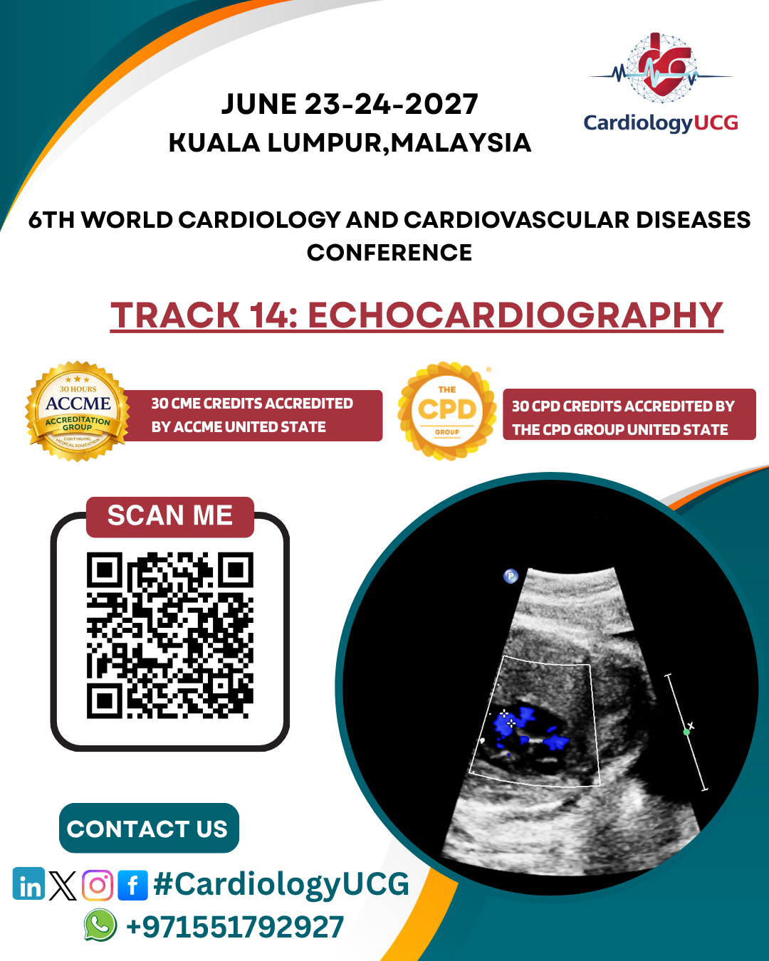
What is echocardiography ?
Echocardiography is a non-invasive imaging technique used to visualize the heart and assess its structure and function. It uses sound waves (ultrasound) to create real-time images of the heart, allowing doctors to evaluate how well the heart is pumping blood, the condition of the heart valves, and the size and movement of the heart chambers.
How Echocardiography Works:
Echocardiography involves the use of a device
called a transducer that emits high-frequency sound waves.
These sound waves travel through the body and bounce off the heart structures,
creating echoes. The echoes are then captured by the transducer and converted
into images displayed on a monitor.
Types of Echocardiography:
1.
Transthoracic
Echocardiography (TTE):
- This
is the most common form of echocardiography, where the transducer is
placed on the chest wall to capture images of the heart. It is typically
done while the patient is lying down.
2.
Transesophageal
Echocardiography (TEE):
- In
this method, a small probe is passed down the esophagus to get closer
images of the heart. It is often used when clearer images are needed or
when TTE is not sufficient. TEE is particularly useful for visualizing
the heart's back structures, such as the left atrium and mitral valve.
3.
Doppler
Echocardiography:
- This
technique measures the movement of blood within the heart and blood
vessels. It helps evaluate blood flow, detect blockages, and assess valve
function. Doppler echocardiography can identify abnormalities in blood
flow, such as regurgitation or stenosis.
4.
2D
Echocardiography:
- Traditional
echocardiography that produces two-dimensional (2D) images of the heart.
This is the most widely used method for evaluating the heart's structure,
chamber sizes, and overall function.
5.
3D
Echocardiography:
- Provides
three-dimensional (3D) images of the heart, allowing for a more
comprehensive view of heart structures and better assessment of complex
heart conditions, such as valve issues.
6.
Stress
Echocardiography:
- This
involves performing an echocardiogram while the patient is undergoing
physical activity or pharmacological stress to evaluate how the heart
responds to exercise or stress. It is used to assess coronary artery
disease, heart function under stress, and to identify areas of the heart
that may not be receiving enough blood.
Uses of Echocardiography:
1.
Evaluating
Heart Function:
Echocardiography is commonly used to assess the overall function of the heart,
including the ejection fraction (the percentage of blood
pumped out of the heart with each beat) and how well the heart chambers are
pumping.
2.
Diagnosing
Heart Disease:
It is crucial in diagnosing various forms of heart disease, such as heart
valve disease (mitral valve prolapse, aortic stenosis, etc.), cardiomyopathy,
pericardial disease, and congenital heart defects.
3.
Monitoring
Heart Failure:
Echocardiography is used to monitor patients with heart failure,
helping doctors track changes in heart size, function, and fluid accumulation.
4.
Detecting
Valve Problems:
It helps identify problems with the heart valves, such as stenosis
(narrowing of the valve), regurgitation (leakage of blood
through a valve), or prolapse (when the valve does not close
properly).
5.
Assessing
Blood Flow:
Doppler echocardiography evaluates the speed and direction of blood flow,
allowing doctors to identify blockages, stenosis, or abnormal blood flow
patterns that could indicate cardiovascular issues.
6.
Congenital
Heart Defects:
Echocardiography is essential in diagnosing congenital heart defects in both
infants and adults, helping to visualize structural abnormalities in the heart
and great vessels.
7.
Monitoring
Post-Surgery Recovery:
After heart surgeries such as valve replacements or coronary artery bypass
grafting (CABG), echocardiography is used to assess the success of the
procedure and monitor for any complications.
Benefits of Echocardiography:
- Non-invasive: No need for
surgery or insertion of instruments into the body.
- Safe: There’s no radiation
involved, making it safe for repeated use.
- Real-time imaging: It
provides live images of the heart, allowing doctors to evaluate heart
function during the exam.
- Quick and accessible: The
procedure is generally quick, and results are available immediately,
making it an essential tool for urgent cardiac assessments.
Limitations of Echocardiography:
- Image quality: In some
patients, particularly those with a high body mass index (BMI) or lung
disease, it can be challenging to obtain clear images.
Technical expertise: The quality of the results depends on the
skills and experience of the technician or cardiologist performing the
procedure