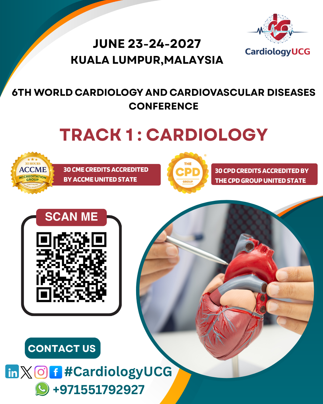
Sub Topics: Coronary Artery Disease, ...
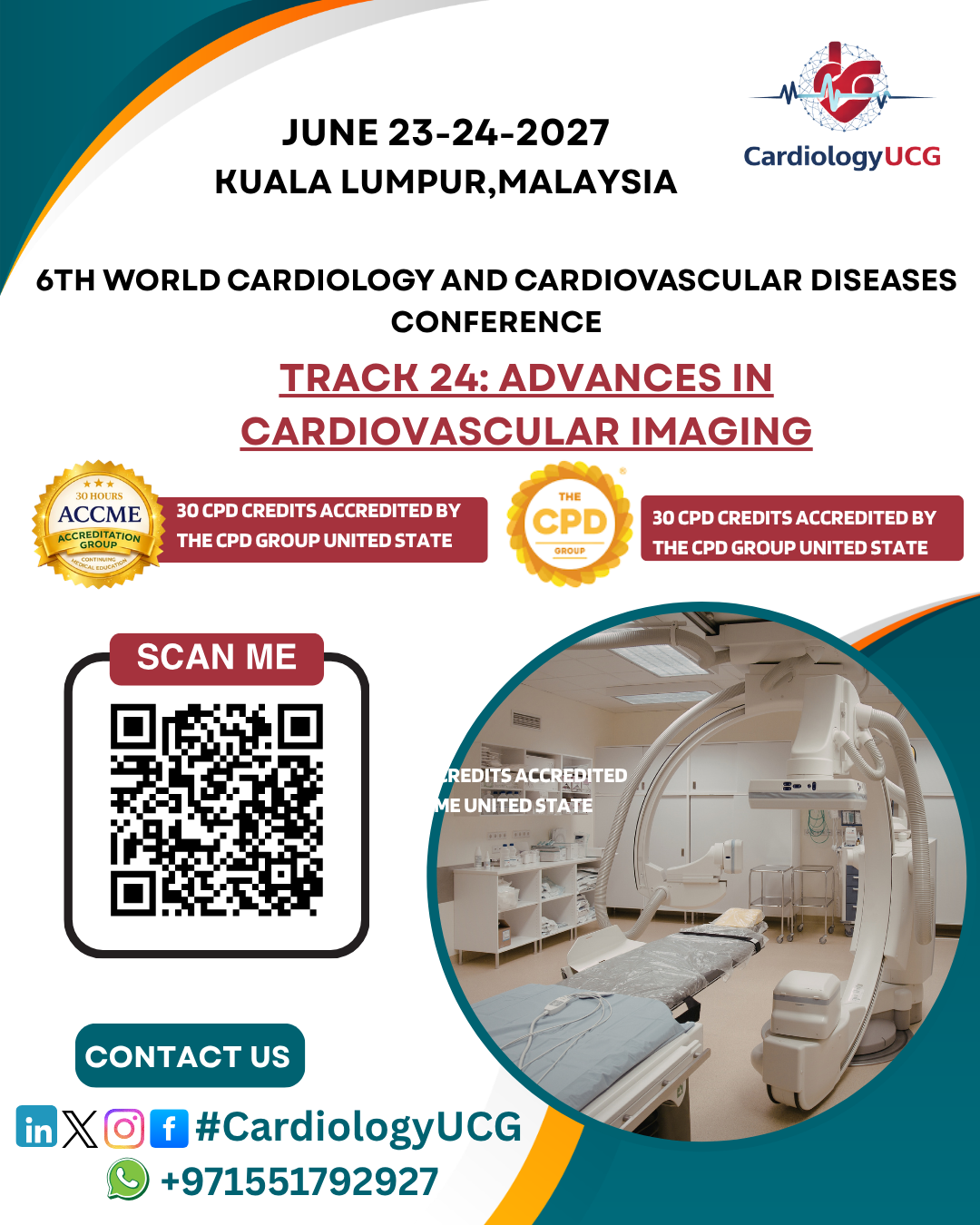
Sub track of advances-in-cardiovascular-imaging : advances-in-cardiovascular-imaging, 3D Echocardiography, Cardiac MRI, Cardiac CT Imaging, Contrast-Enhanced Ultrasound, Positron Emission Tomography (PET), Coronary CT Angiography, Magnetic Resonance Angiography (MRA), Myocardial Strain Imaging
Advances in Cardiovascular Imaging refer to the innovative
techniques and technologies developed to improve the ability to visualize and
assess the heart and blood vessels. These advancements have significantly
enhanced the accuracy of diagnosing cardiovascular diseases (CVDs), planning
interventions, and monitoring treatment responses.
Here are some key areas of advances in cardiovascular imaging:
Echocardiography, a non-invasive imaging technique using sound waves, has
evolved with improvements in image resolution and techniques. 3D
echocardiography allows for more detailed visualization of heart
chambers and valves, enhancing the diagnosis of conditions like valvular heart
disease and congenital heart defects.
This advanced form of echocardiography creates three-dimensional images of
the heart, providing better visualization and more precise measurements of
cardiac structures. It is especially useful for assessing heart valves,
ventricular function, and congenital heart diseases.
Cardiac MRI has become a cornerstone in evaluating heart function and
detecting conditions like myocarditis, cardiomyopathy, and coronary artery
disease. Advances in late gadolinium enhancement (LGE) imaging
allow for better detection of heart tissue damage, while Cine MRI
provides moving images of the heart’s function.
Cardiac CT uses X-ray technology to obtain detailed cross-sectional images
of the heart. Advances in coronary CT angiography enable
precise imaging of the coronary arteries, helping in the diagnosis of coronary
artery disease (CAD), aneurysms, and other vascular conditions.
This technique uses ultrasound contrast agents to enhance the quality of imaging,
particularly in detecting blood flow and identifying conditions like myocardial
ischemia and heart valve abnormalities. It provides real-time, dynamic images
of the heart's structure and function.
PET imaging is used to assess blood flow and metabolism in the heart. Recent
advances in PET myocardial perfusion imaging have helped in
identifying areas of the heart that are at risk of ischemia and can guide
decisions for interventions like coronary artery bypass surgery (CABG).
Coronary CT angiography allows for non-invasive imaging of coronary
arteries, detecting blockages, aneurysms, and coronary artery disease (CAD). It
is now a standard diagnostic tool for evaluating heart disease, especially in
patients with intermediate risk.
MRA is used to visualize blood vessels, and it is particularly useful for
detecting vascular anomalies, aneurysms, and stenosis. It is a non-invasive
alternative to catheter angiography and offers clear, detailed images of the
arteries.
Strain imaging assesses the deformation (strain) of the heart muscle during
contraction. This technique, particularly with speckle-tracking
echocardiography, is useful in detecting subtle changes in heart
function, often before traditional imaging can identify problems, especially in
heart failure and cardiomyopathy.
Advancements in imaging technologies allow for higher resolution
scans, leading to clearer images of small vessels and heart tissues.
These techniques are essential for detecting early stages of heart disease,
such as the formation of plaques in coronary arteries.